“Mammalian eyes. Ahh, I see”.
This common statement, linking – as it does – understanding with sight, is an excellent example of how important vision is to Humankind.
We are the most visually orientated and vision dependent of all the mammals.
However, nearly all mammals use sight to some degree. For many it is one of the primary senses, one of their main ways of perceiving the world around them.
It is not surprising then, that the mammalian eye and those parts of the brain associated with interpreting the data gathered by the eye are quite well developed.
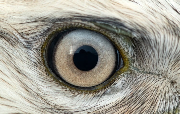
Mammalian eyes, like all other eyes, is a device for collecting and focusing light rays; for distinguishing between different wavelengths and intensities; and for converting that information into electrical impulses that are sent to the brain.
The Most Important Parts Of Mammalian Eyes
The Eyelids
Mammalian eyes have two eyelids, one upper and one lower eyelid, of which the upper is more moveable. Birds and some reptiles have 3 eyelids.
Our eyelids are used in cleaning and protecting the eye. They can be closed to protect the eye from physical assault or from excess light. Blinking helps spread lachrymal fluid (tears) across the front of the cornea, clearing away dust and preventing the cornea from becoming dried and cracked.
The Cornea
This is the protective covering on the outer surface of the eye.
The Anterior Chamber
This is the space immediately behind the cornea and leading to the iris and the lens. It is filled with a fluid called the aqueous humour.
The Iris
This is a muscularly operated diaphragm which controls the amount of light entering the eye. It is coloured and it is this that gives the eye its colour. At the centre of the iris is the variable hole through which the light actually passes on its way into the eye, this is called the pupil.
The Lens
This is a transparent convex or ‘lens’ shaped body with a harder outer layer and a softer inner layer. It serves to focus the light on the retina. Its shape can be altered by means of the ciliary muscles which are attached to the eye by means of the zonular fibres.
The Posterior Chamber
This is the bulk of the eye and is the space behind the lens and between the lens and the retina. It is filled with a clear jelly-like substance called the vitreous humour.
The Retina
This is the inner light receptive part of the eye. It is covered in special ‘photoreceptive’ cells called rods and cones. Towards the centre of the retina is an area called the fovea centralis, which has a greater density of rods and cones. This is the area of greatest visual acuity, i.e. sharpest , clearest detection of objects.
The Optic Nerve
This is a bundle of nerve fibres which carry messages from the eye to the relevant parts of the brain and vice-versa. There is a small blind spot where the optic nerve meets the retina.
The Sclera
Sometimes called the Sclerotic, this is a tough collagen fibre layer which surrounds the whole inner part of the eye (that part not covered by the cornea). It supports and protects the eye as a whole.
Light Detection in Mammalian Eyes
Light is an electromagnetic radiation that travels as a compression wave through a few substances, including air. Electromagnetic radiation has a number of defining properties.
The one that concerns us most is wavelength. This is the distance between successive troughs or crests in the wave. In nature there are a huge range of wavelengths but only a few are used by mammals, mostly those between 350 and 750 nanometres.
One nanometre is 10-6 of a metre, which is one 1,000,000th of a metre, or not very long. Different species of mammals perceive different wavelengths more efficiently than others.
The light detecting organ in the eye is the ‘retina’ which lines the inside of the eye and is covered in special receptors. These receptors absorb the light that hits and then generate an electrical impulse which travels down the ‘optic nerve’ to the brain.
The brain receives thousands of these small messages every millisecond that you eyes are open and it uses them to build up the constantly changing image of the world that you live from.
There are two sorts of receptors in the mammal eye. They are called ‘rods’ and ‘cones’. Rods are better for night vision because they are sensitive to small quantities of light. Some nocturnal animals such as opossums have only rods in their retina which gives them excellent night vision.
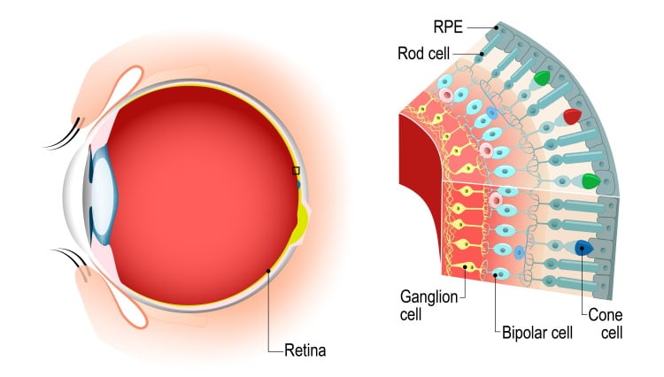
However as it is the cones that allow us to distinguish between different colours of light, they miss out on a lot of the beauty of the world. Also there can be as many as 150 rods connected to a single nerve cell, this increases its sensitivity but reduces its definition, i.e. things look blurry.
Cones can be as few as one or two per nerve cell. This works the opposite way making them less sensitive but more accurate for detail. Which is good during the daylight. The number of receptors per square millimetre determines the degree of visual acuity an animal has – the more receptors the higher its ability to distinguish individual objects at a distance.
We have about 200,000 receptors per mm2 which is pretty good for a mammal but does not really compare well with a Buzzard which has an incredible 1,000,000 receptors per mm2.
Cones come in different flavours depending on which pigment they contain, different pigments detect different colours. Most humans have 3 pigments: red, green and blue. Some people lack one of the pigments and this gives them a sort of colour blindness.
Variations in Mammalian Eyes
Other mammals do not see colour as well as we do, though most mammals see some colour. They may have only one or two pigments, or they may have different pigments to us altogether. Sometimes not every member of a species has the same pigments.
Squirrel Monkeys have two or three pigments in their eyes, but they have five different pigments potentially available. If we call the pigments 1,2,3,4 and 5 we can see that one monkey could have pigments 1 and 2, while another could have pigments 3, 4 and 5 .
This would mean both monkeys would see the world very differently. It would make the evolution of a language to describe colour very difficult.
Most mammals have a mixture of rods and cones in their retinas. Most mammals also experience a variety of light conditions, night, dawn, day and dusk. Therefore it makes sense to have a mechanism to control the amount of light that enters the eye.
A way of ensuring as much light as possible reaches the retina under dull or dark conditions, but capable of controlling the influx of light – so that on bright days the sensitive retina does not get damaged by an excessive of light.
This mechanism is called the ‘iris’, and it exists just outside of the lens. It is a circular sheet of muscle and through muscular control we can make its diametre larger or smaller… and thus control the amount of light that enters the eye.
Look into a friends eyes and see the size of their ‘pupil’.
This is the hole in the centre of the iris through which the light passes. Once you have had a good look at the size of their pupil, ask them to close their eyes and to put their hands over their eyes for a minute.
Then look into their eyes again. Not only should you see that the pupil is larger, but you should be able to see it shrink in size as their eyes adjust to normal daylight again. This is an involuntary muscular action, meaning you do not choose to open or close your iris, but your body is automatically looking after itself by responding to changes in light density.
Nocturnal animals such as cats can open their irises far wider than we can, giving them much better night vision than us. Primarily nocturnal animals also have a special organ called a ‘tapetum’.
This is a layer of cells in the ‘choroid’ which lies just behind the retina. It reflects light back through the light sensitive area of the retina, thus giving the rods and cones a second chance to detect the light. It is this tapetum which gives animals like foxes eyes that seem to shine at night in the headlights of your car.
Some animals, such as man, have dual purpose eyes with a special area called a ‘fovea’ – which is rich in cones while the rest of the retina is rich in rods. This allows us to see well in daylight but also to see reasonably well at dusk and dawn.
The Field Of Vision in Mammalian Eyes
The positioning of the eyes on the head is important because it controls the animals ‘field of vision’. Which is how much of the world it can see without moving its head.
For humans this is about 180 degrees of a circle, if measured laterally (from side to side) as it normally is.
For a horse it is about 215 degrees. This means that horses and similarly designed animals can see more of the world at any given moment than a human or a cat. For a horse it is important to be able to see a predator before it gets close. It doesn’t need to see it clearly or to know exactly how close it is, just to know the minute it comes into vision.
For carnivores, and omnivores (part-time predators) it is more important to be able to make accurate judgments concerning another animal’s distance and movement.
To do this we need binocular vision. In other words we need to see the object with both eyes. This means the ‘fields of vision’ of both eyes have to overlap – and thus the eyes have to be more to the front of the head. Inevitably this reduces our total field of vision, but makes hunting possible.
Binocular vision works because our eyes are still slightly apart, meaning that the image each eye delivers to the brain is slightly different. Our brains use this difference to build up a 3D image of the world.
This allows us to make much better judgments of how far away things are and how fast they are moving. The world as seen through through the single eyes of a herbivore is much flatter.
Most mammals have some binocular vision. For animals like a horse the overlap is only of the peripheral vision and represents a small percentage of its view of the world. Truly binocular animals like primates do most of their seeing binocularly and have only a few degrees of monocular vision around the edges of our sight.
In mankind, binocular vision is about 140 degrees out of a total of about 180 degrees.
Of course, all this seeing only works if the image is clear (in focus). A blurred image conveys much less information, or less accurate information, to our brains.
To keep the image focused properly, we need the incoming light to be focused on the retina – and for this we have a lens in our eyes. The lens sits immediately behind the iris at the front of the eye. A lens with a fixed shape would focus objects at different depths in the eye, depending on how far away they are.
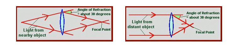
Objects that are further away naturally focus closer to the lens. This is because all the lens does is bend (refract) the light by a certain amount. The angle at which the light rays hit the lens determines the angle at which they leave the other side of it.
Light beams from a distant object will be travelling nearly parallel to each other, while those from a nearer object will be diverging more (see diagram).
The image of an object is focused when all the light rays coming through the lens from the object pass meet a single point. This is called the ‘point of focus’. When this point of focus lands squarely on the retina, the object will be seen clearly.
There are two ways nature gets around the problem of having objects from different distances focusing at different depths within the eye. The first is seen in animals like the coleoid cephalopods. In these animals, whose eyes are in many ways as complex as our own, the lens can be moved a little backwards and forwards in order to facilitate better focus
Humans however use a different method.
Nature’s second solution is to have a flexible lens and muscles attached to it. By changing the shape of the lens you change the angle of refraction of the light passing through the lens and thus move the point of focus closer to or further away from the lens.
This means you can bring distant or close objects into sharp focus at the twitch of a muscle. These systems are useful for animals that need accurate images of different parts of the world all the time, such as hunters. Other mammals, like horses, can adjust the shape of their lenses as well, but not to the extent that humans can.
What Next?
Well, I hope this has been an interesting look into the workings of the mammalian eye!
Perhaps now you’d like to learn about the mammal digestive system.